


Retinopathy of premature babies
(ROP – retinopathy of prematurity)
Premature babies with low body weight may develop proliferative retinopathy, a disease of the fundus due to insufficient development, or retinopathy of prematurity. Retinal blood vessels begin to develop in the mother’s womb and continue to develop after birth, i.e. until the end of the first month after birth. The retina is then particularly sensitive and various damages can occur, which is especially evident in premature babies.
There are active and scarring (old, stable) forms of this disease. The severity of the change is determined based on: the localization of the change on the fundus, the extension of the change, the advanced stage and associated eye diseases. As a result of insufficient development, ischemic, i.e. bloodless zones appear on the fundus. These are fields with increased pathological vascularization that try to compensate for the lack of blood vessels, as well as fields with scar formation. Bleeding in the eye, the occurrence of ablation and finally, irreversible blindness can occur as more serious complications.
Signs of the disease cannot be noticed by parents, nor is the baby capable of pointing them out.
Screening and timely examination are of crucial importance for the detection and early treatment of this disease.

SCREENING
All babies who are born from high-risk pregnancies, twin pregnancies, born before the 37th week of gestation or with a weight of less than 2 kg should be examined immediately at birth. Subsequent follow-ups should be performed two weeks after the first examination, until the retina has fully developed. In the sixth or seventh month after birth, another examination should be done.
TREATMENT
Depending on the findings, there are several types of treatment, which fit according to the indications. The use of anti-VEGF therapy, the use of lasers and, finally, the possibility of performing vitreoretinal surgery (eye fundus surgery) also come into consideration. It should be noted again that everything depends on the stage and signs of the disease, and that it is very important to treat everything at the right time. For all the above reasons, only timely screening can help.
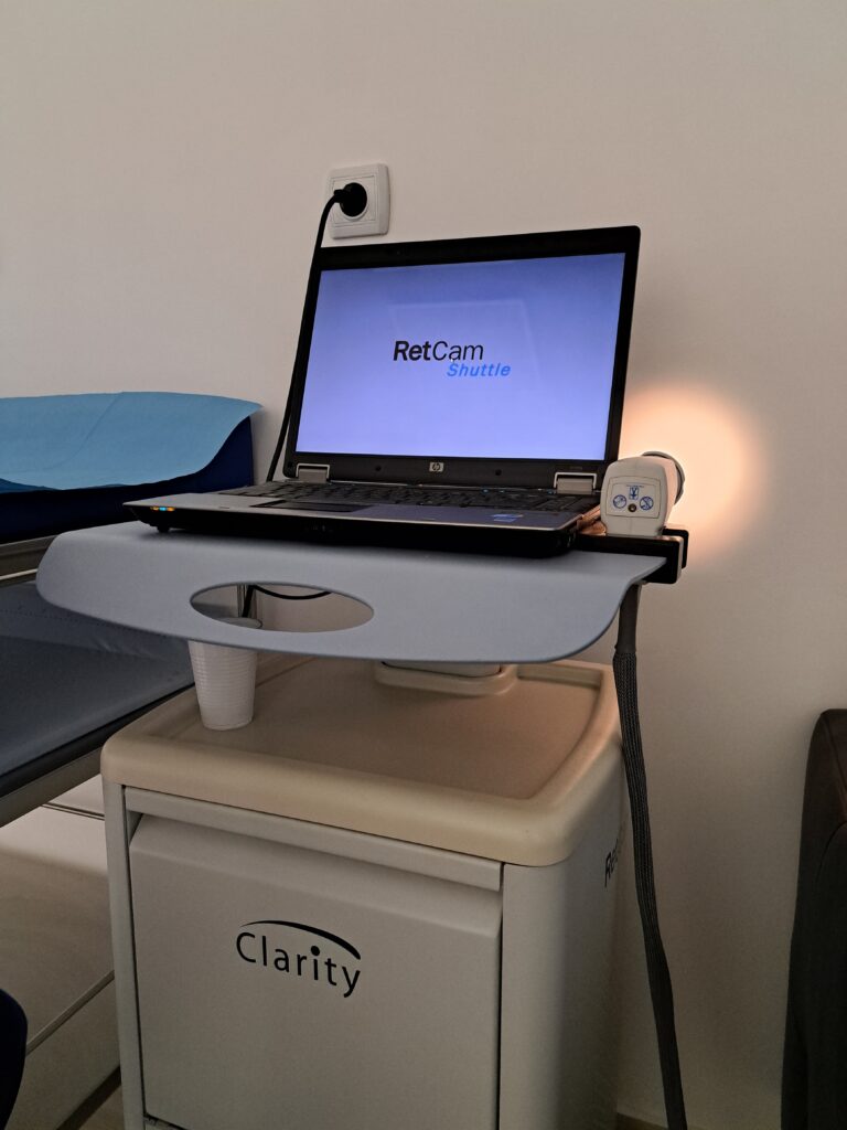
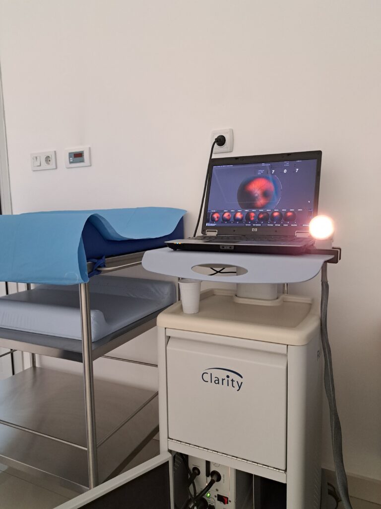
RetCam – THE MOST MODERN CAMERA FOR IMAGING THE FUNDUS OF BABIES
RetCam examination is a basic examination for all babies who are suspected of underdevelopment of the eye and vision in general. With all premature babies, babies from high-risk or twin pregnancies, as well as with some infections during pregnancy, it is necessary to examine the fundus immediately after birth.
The examination is performed by a specially formed, expert team for children’s ophthalmology, which represents a combination of experience and youth. With the RetCam, the recording of the fundus in children has become significantly more comfortable, the baby does not feel any discomfort, its cooperation is not required, and what is most important, the findings are saved as video materials and images. The recorded documents can be interpreted in detail by doctors after the examination and compared with previous results. The camera is specially designed so that it can record the entire fundus and from hard-to-reach angles, without coming into contact with the surface of the eye. In this way, the possibility of any injury is minimized and the procedure is extremely safe.
“Maja Clinic” is the first special hospital for ophthalmology in Serbia that provides imaging with the RetCam device. Until the appearance of this camera on our market, the possibility of printing and saving findings, the possibility of comparing data and analyzing changes, were not possible. RetCam has brought many advantages and therefore holds the title of the most modern device (camera) for imaging the fundus of babies.
What does the RetCam examination look like at the “Maja Clinic”?
In order for parents and children to have comfort during their visit, the “Maja Clinic” has provided special premises for filming. When parents come with their child, the anterior segment of the eye is first examined, followed by dilation of pupils. After about an hour, when the pupils are sufficiently dilated, the recording is performed with the RetCam device. It does not last longer than 10 minutes. At the end of the examination, parents receive the doctor’s opinion, as well as the findings in printed and electronic format.
The advantages of this way of examination are:
- Comfort for children and parents
- Shorter examination time
- Portable image record of the examination in electronic and printed form
- Timely treatment
Babies and children at an early age, when their vision is developing the most, cannot and do not know how to ask for help from adults. The team of experienced doctors at the “Maja Clinic” wants to pay full attention to the youngest at these important moments and contribute to their healthy vision development. Doctors who have experience in working with children, along with young and promising ophthalmologists who bring innovations in the form of implementation of advanced technologies, represent a perfect combination for the healthy development of the vision of our youngest patients.