


Visual Field Test
A visual field test is an eye examination that can detect dysfunction in central and peripheral vision which may be caused by various medical conditions such as glaucoma, stroke, brain tumours or other neurological defects. Visual field testing can be performed clinically by keeping the subject’s gaze fixed while presenting objects at various places within their visual field. Simple manual equipment can be used such as in the tangent screen test of the Amsler grid. When dedicated machinery is used it is called a perimeter.
Automated (computerised) perimetry
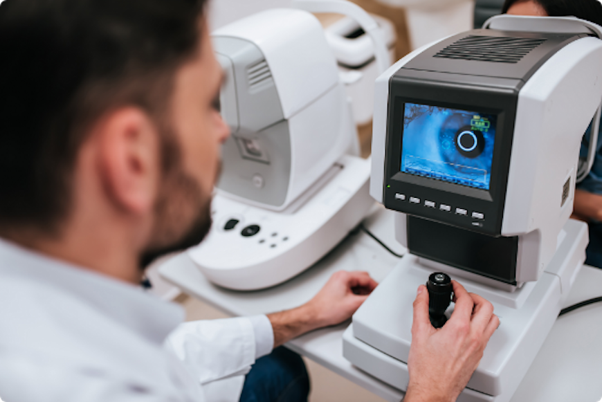
Uses a mobile stimulus moved by a perimetry machine. The patient indicates whether he sees the light by pushing a button. The use of a white background and lights of incremental brightness is called ”white-to-white” perimetry. This type of perimetry is the most commonly used in clinical practice, and in research trials where loss of visual field must be measured. However, the sensitivity of white-to-white perimetry is low, and the variability is relatively high: as many as 25-50% of the photoreceptor cells may be lost before changes in visual field acuity are detected.
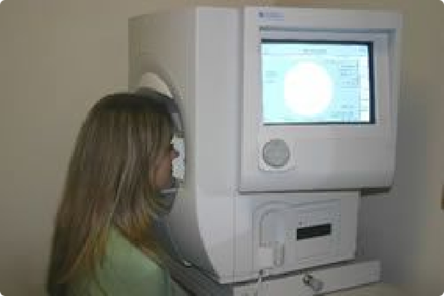
This method is commonly used for early detection of blind spots. The patient sits in front of an (artificial) small concave dome in small machine with a target in the centre. The chin rests on the machine and the eye that is not being tested is covered. A button is given to the patient to be used during the exam. The patient is set in the front of the dome and asked to focus on the target at the centre. A computer then shines lights on the inside dome and the patient clicks the button whenever a light is seen. The computer then automatically maps and calculates the patient’s visual field.
Other types of primetry are: Tangent screen, Goldmann perimeter, Microperimeter.
Methods of stimulus presentation:
Static perimetry (it is used for rapid screening and follow up of the diseases involving defects such as scotomas, loss of peripheral vision and more subtile vision loss and is important in the screening, diagnosing, and monitoring of various eye, retinal, optic nerve and brain disorders)
Kinetic perimetry (is useful for mapping visual field sensitivity boundaries, and may be a good alternative for patients that have difficulty with automated perimetry).
The most important use of this method is:
Glaucoma – Detection and monitoring. The patient is often not aware of this disease, but there are some typically defects of the visual field which can be an important signal that something is happening. It is very important to repeat this examination every 3-6 months, especially after the glaucoma operation.
Neurological defects – It is a simple but important method for detection a localization of the neurological defect.